With weight recovery, plasma neurofilament light chain levels fell.
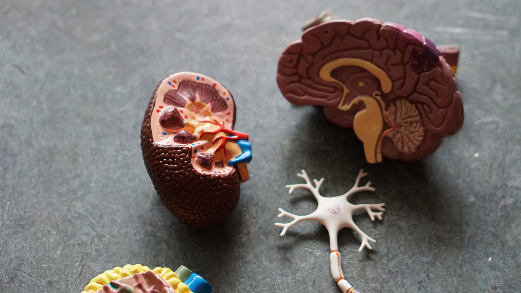
A number of structural brain changes have been identified in patients with AN, including reductions in grey or white matter, as well as enlarged sulci and ventricles. Swedish scientists have examined a potential blood marker of these brain effects: increased plasma neurofilament light chain (NfL) concentrations. This blood-based biomarker (an indication of neuronal damage) is often used to follow disease progression in conditions such as Parkinson’s disease or Alzheimer’s disease.
Molecular biologist Ida A. K. Nilsson and a team at the Karolinska Institute, Stockholm, hypothesized that neuronal injury and/or degeneration is involved in the pathophysiology of AN. To test this, they measured levels of NfL in a small study of women with AN (n=12), women with AN who were weight-recovered (n=11), and normal-weight, age-matched, controls (n=12) (Translational Psychiatry. 2019; 9:180). They also sought to learn if such changes are due to degeneration of brain cells or merely to changes in fluid levels.
The team found significantly higher plasma NfL levels among the women with AN, compared to the normal-weight women with and without a history of AN. NfL levels were negatively associated with body mass index in all three groups. The results suggested that the light chain levels might normalize with weight recovery. Other studies of different neuronal change markers have shown varying results after weight is regained, and the authors could only speculate about the origin of the neuronal injury tied to elevated Nfl levels. They did note that it is possible that the blood-brain barrier becomes more permeable in conditions of starvation, such as AN, which would allow more NfL to leak into the circulation. Further study is needed to better pinpoint areas of the brain that are affected, and long-term effects among AN patients.
